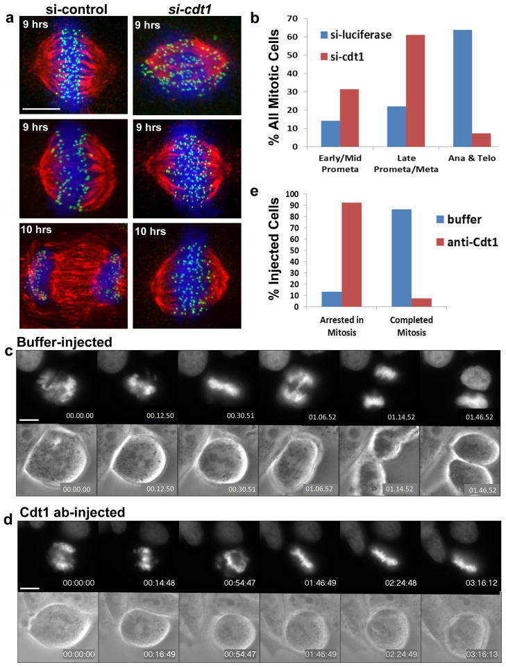
Figure 2. G2-specific Cdt1 inhibition induces mitotic arrest.
(a) HeLa cells synchronized in early S phase were released into control (luciferase) or cdt1 siRNA for either 9 hrs (top and middle panels) or 10 hrs (bottom panels) followed by fixation and staining with DAPI to label chromosomes (blue), anti-tubulin antibody to label MTs (red), and anti-Knl1 antibody to label kinetochores (green). (b) Quantification of the results at 10 hr in a by mitotic stage; n = 1500 cells. (c–e) HeLa cells stably expressing GFP-histone H2B were injected with control buffer (c, n=15) or anti-Cdt1 antibody (d, n=27). Selected frames of GFP-histone (top panels) and phase contrast images (bottom panels) are shown. (e) Quantification of the results from the microinjection experiments in c and d. Scale bars = 5 μm. See also Supplementary Movies S1–S4.

