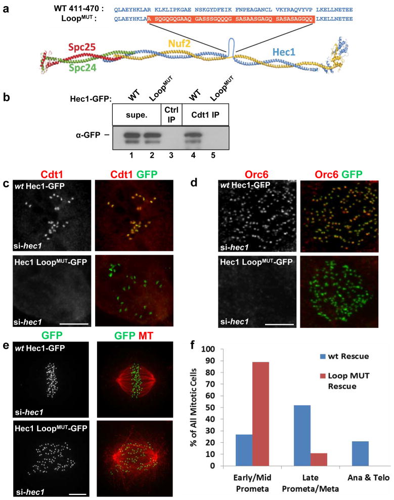
Figure 5. Cdt1 targeting to kinetochores depends on the flexible loop region of Hec1.
(a) Diagram of the Ndc80 complex showing the loop region and the construction of the Hec1 loop replacement mutant,“Hec1 LoopMUT”, (adapted from Ciferri et al., 200835). (b) Endogenous Cdt1 was immunoprecipitated from lysates of asynchronously growing HeLa cells transfected with Hec1-GFP plasmids; “Ctrl IP” indicates the use of normal mouse serum as a control. Hec1-GFP in the bound (“Cdt1 IP”) and unbound (“Supe.”) fractions was detected with anti-GFP antibody. (c) PTK2 cells were treated with hec1 siRNA followed by transfection with either wt Hec1-GFP or Hec1 LoopMUT-GFP constructs. The cells were treated with nocodazole then fixed and stained using anti-Cdt1 antibody and anti-GFP antibody. (d) As in c except that HeLa cells were stained with anti-Orc6 and anti-GFP antibodies. (e) As in c except that HeLa cells were stained with anti-tubulin antibody to mark MTs and anti-GFP antibody to mark Hec1-GFP at kinetochores. (f) Quantification of mitotic stages in Hec1-depleted cells expressing ectopic wt Hec1-GFP or Hec1 LoopMUT-GFP stained with DAPI; n = 125 GFP-expressing cells. Scale bars = 5 μm. See also Supplementary Figure S8 and Supplementary Movies S5 and S6.

