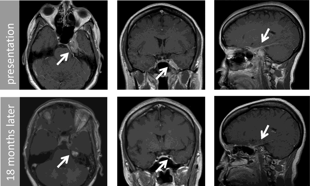
Figure 3.
Post contrast T1 MRI of brain axial (left column), coronal (center column) and saggital (right column) images from patient 2. These demonstrate an extra-axial lesion involving left middle and anterior cranial fossa, petrous apex, cavernous sinus, cerebellopontine angle and tentorium. Top row shows initial imaging and bottom row shows 18 month follow up.

