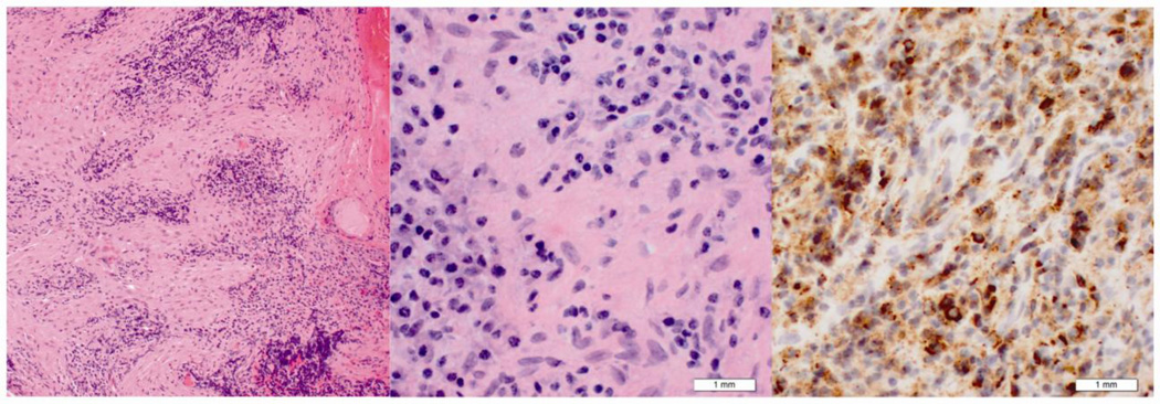
Figure 4.
Histologic features of middle cranial fossa biopsy from patient two. Hematoxylin and eosin stain at low (10× objective lens) (left) and high (40× objective lens) (middle) power. These demonstrate a mixed inflammatory infiltrate and fibrosis. IgG4 immunohistochemical stain (right) demonstrates multiple immunoreactive plasma cells.

