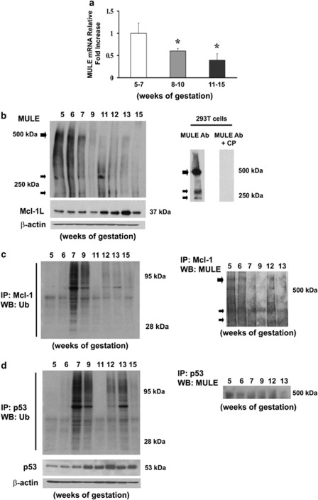
Figure 1.
Expression and association of MULE, Mcl-1 and p53 during human placental development (5–7 weeks, n=19; 8–10 weeks, n=14; 11–15 weeks, n=24). (a) MULE mRNA expression during placental development assessed by real-time PCR (5–7 weeks, n=7; 8–10 weeks, n=6; 11–15 weeks, n=12). Values are mean±S.E.M., *P<0.05. (b) Representative immunoblots for MULE (upper left panel) and Mcl-1 (lower left panel) protein expression during placenta development and MULE antibody validation (right panel) assessed by MULE immunoblot using 293T cells in the presence or absence of competing peptide (CP). (c) Representative immunoblots for Mcl-1 ubiquitination (left panel) and MULE/Mcl-1 association (right panel) during placental development. (d) Representative immunoblots for p53 protein expression (left lower panel), ubiquitination (left upper panel) and MULE/p53 association (right panel) during placental development. (b and d) Actin immunoblots demonstrates equal protein loading (5–7 weeks, n=12; 8–10 weeks, n=8; 11–15 weeks, n=12)

