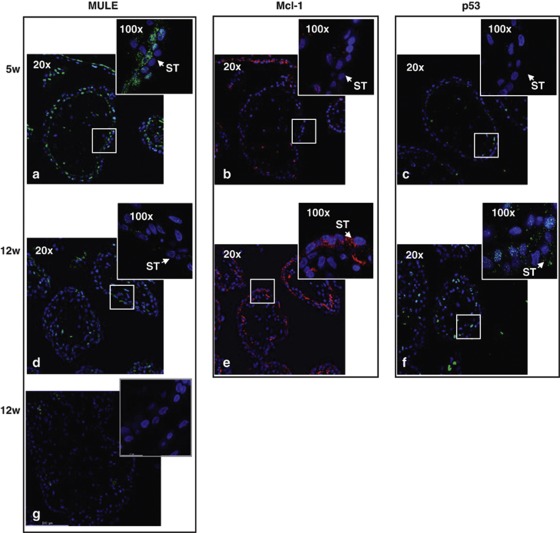
Figure 2.
MULE, Mcl-1 and p53 spatial localization in first trimester human placental sections. MULE (a and d), Mcl-1 (b and e) and p53 (c and f) spatial localization at 5 and 12 weeks of gestation assessed by immunofluorescent staining (5–7 weeks, n=7; 8–10 weeks, n=6; 11–15 weeks, n=12). Cell nuclei are showed in blue by DAPI signal. ST, syncytiotrophoblast cells. Control IgG (g). Original magnifications, × 20; insets, × 100

