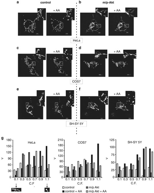
Figure 4.
Akt preserves the mitochondrial integrity in an IP3R type III-dependent way. (a–f) The mitochondrial network of HeLa (a and b), COS7 (c and d) and SH-SY 5Y cells (e and f) was analyzed using confocal microscopy, 36 h post-transfection, before and after treatment with 80 μM AA for 20 min. Cells were co-transfected in a 3 : 1 ratio with an empty vector and mtGFP (left panels: control) or m/p-Akt and mtGFP (right panels: m/p-Akt). Greater magnification of the mitochondrial three-dimensional structure is presented in the insets. (g) Frequency distribution of mitochondrial fragmentation: C.F. was analyzed to represent the state of each single mitochondrial object (i.e., C.F.=0.1 ‘elongated' mitochondrion, considered not fragmented; C.F.=1.1 ‘circular' mitochondrion, considered fragmented). ν, frequency (number of objects)

