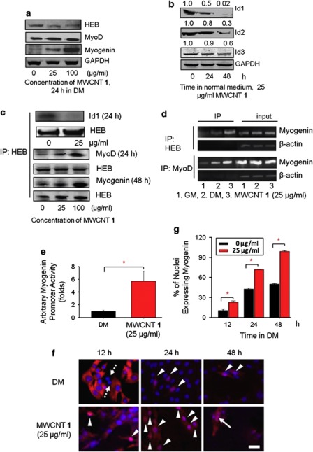
Figure 2.
MWCNT 1 positively regulates functions of MyoD and myogenin by inhibiting expression of Id proteins. (a) Effects of MWCNT 1 on the expression of MyoD, myogenin and HEB analyzed by western blots. (b) MWCNT 1 decreased the expression of Id proteins. (c) MWCNT 1 caused a decrease in HEB–Id complex formation and an increase in HEB–MyoD and HEB–myogenin. Protein complex formations were analyzed by co-immunoprecipitation with anti-HEB antibody followed by probing the co-precipitated proteins with different antibodies after MWCNT 1 treatment. (d) MWCNT 1 recruiting HEB–MyoD complexes to E-box motifs in the promoter of myogenin gene assayed by chromatin immuoprecipitation (ChIP) with anti-HEB and anti-MyoD antibodies. Total DNA before immunoprecipitation was used as input controls and β-actin was used as a control for specificity (Supplementary Information for details). (e) Myogenin promoter activity enhanced by MWCNT 1. C2C12 was transfected with constructs containing firefly luciferase gene driven by myogenin promoter and the luminescence intensity was measured after treatment for 48 h. The luminescence intensity in DM was normalized to 1.0. (f) Myogenin expression and distribution in C2C12 cultured in DM or MWCNT 1 for various times. Myogenin-negative cells were indicated by arrows with dotted line and cells with nuclear translocation of myogenin were shown with arrow heads. Fusion of myogenin-positive nuclei was indicated by arrows with solid line. Immumnocytochemistry was performed with anti-myogenin antibody and Cy3-conjugated secondary antibody. (scale bar: 50 μm). (g) Quantitative analysis of nuclei expressing myogenin at different time points (*P<0.05)

