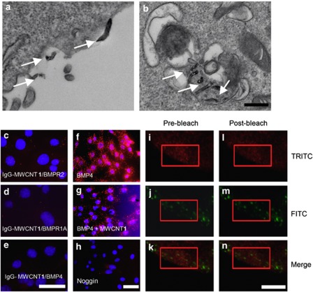
Figure 5.
MWCNT 1 blocks phosphorylation of BMPR1A by binding to BMPR2. (a and b) TEM images show MWCNT 1s (arrows) bound to cell membranes or in endosomes. C2C12 cells were incubated with MWCNT 1 (25 μg/ml) for 2 h before fixation. Scale bar, 100 nm. (c–h) MWCNT 1 has a close proximity with BMPR2 and interferes with BMPR1A–BMPR2 complex formation assayed by in situ PLA. In (c-e) MWCNT 1 was conjugated with mouse IgG, and protein pairs between IgG with rabbit BMPR2 (c), BMPR1A (d) and BMP4 (e) antibodies were determined. PLA signals were shown in red, and nuclei were stained blue by DAPI. (f–h) BMPR1A–BMPR2 complex formation was assayed by in situ PLA after addition of BMP4 (f), BMP4 with MWCNT 1 (g) and BMP4 with noggin (h). C2C12 cells were treated with MWCNT 1 (25 μg/ml) or noggin (200 ng/ml) for 2 h followed by BMP4 (25 ng/ml) treatment for 1 h before fixation. Scale bars, 50 μm. (i–n) MWCNT 1 (labeled by FITC) closely associates with BMPR2 (labeled by TRITC) as detected by FRET. A region in the red rectangle was photobleached at 543 nm (TRITC channel) for 50 exposures and images were taken in FITC (488 nm) and TRITC (543 nm) channels before and after bleaching. Scale bars, 50 μm

