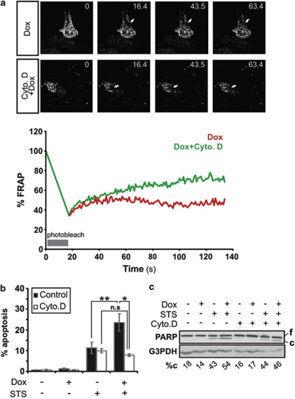
Figure 2.
Cytoskeleton destabilization prevents the enhancement of apoptosis by p57KIP2. (a) FRAP analysis of HeLa-p57KIP2 cells treated with or without cytochalsin D for 3 h. Values represent the % fluorescence recovery over time of actin-GFP after bleaching. Arrows indicate the photobleached area. (b and c) HeLa-p57KIP2 cells were treated with cytochalsin D for 1 h, followed by treatment with STS for 3 h. (b) Apoptotic nuclear morphology was quantified after Hoechst staining and expressed as a percentage of the total cells counted. (c) PARP cleavage was assessed by immunoblotting. G3PDH was used as a loading control. Full length (f) and cleaved (c) PARP was analyzed by densitometry and cleaved PARP was expressed as a % of total PARP (%c)

