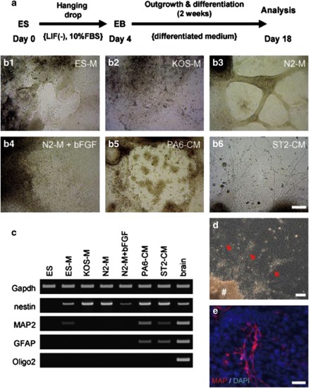
Figure 1.
Directed neural differentiation of ES cells by stromal cells. (a) Schedule of differentiation. Following EB formation, EBs were cultured with various differentiated media (see Table 1) for 2 weeks. (b) Differentiation of EBs was induced using various media, as listed in Table 1. The photographs indicate EB outgrowths after 2 weeks of cultivation: (b-1) ES-M, (b-2) KOS-M, (b-3) N2-M, (b-4) N2-M+bFGF, (b-5) PA6-CM, (b-6) ST2-CM. Scale bar=100 μm. (c) Analysis of expressions neural markers (nestin, MAP2, GFAP, Oligo2) in EB outgrowths after cultivation in various differentiation media for 2 weeks. Samples from adult mouse brain tissues served as a positive control. (d) EB outgrowths cultured with ST2-CM possessed neurite-like structures. The symbol ‘#' and red arrows indicate EB body and neurite-like extension, respectively. Scale bar=100 μm. (e) The neurite-like extensions were immuno-positive for MAP2. Scale bar=20 μm

