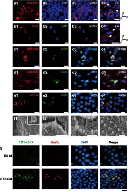
Figure 4.
Stereocilia-like structures formed in HC-like cells. (a–f) EB outgrowths cultured with ST2-CM for 2 weeks were examined with phalloidin staining and immunostaining for Espin, Brn3c, acetylated tubulin as well as with a SEM. (a) Some cells showed phalloidin-labeled protrusions, highly reminiscent of actin-rich hair bundles (arrow). Scale bars=10 μm. (b) Espin-immuno-positive cells were detected, and the immuno-positivity was found in protrusions at the cell rim (arrow). Scale bars=10 μm. (c) Espin-immuno-positive cells (c-2, green) were simultaneously stained for phalloidin (c-1, red). Scale bars=10 μm. (d) Brn3c-immuno-positive cells (d-2, green) were simultaneously stained for phalloidin (d-1, red). Scale bars=10 μm. (e) Acetylated-tubulin-immuno-positivity (e-1, red) was found in Brn3c-immuno-positive cells (e-2, green). Scale bars=10 μm. (f) Distinct stereocilia-like structures were confirmed by SEM analysis. Shown are undifferentiated ES cells (f-1, low magnification in inset), ES cells differentiated with ST2-CM (f-2 and f-3), and inner ear HCs from the organ of Corti of postnatal mice (day 1) (f-4). Scale bars=3 μm (f-1), 2 μm (f-2), 3 μm (f-3), 5 μm (f-4). (g) The mechanotransduction channel function was examined based on rapid permeation of FM1-43FX dye (green), then immunostaining with Brn3c (red) was performed. Scale bars=20 μm

