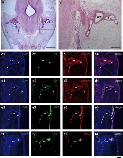
Figure 6.
Transplantation of cells from EB outgrowths containing HC-like cells into chick embryos. (a) Graft cells were transplanted into the otic vesicle of chicken embryos. Three days after injection, normal morphological processes were confirmed by HE staining. Scale bar=1 mm. (b) Enlarged image of area in (a) marked by dotted line. Scale bar=150 μm. (c) No GFP-immuno-positive cells (c-2, green) were found in sections of the inner ear transplanted with undifferentiated ES cells. High phalloidin-positivity was observed in the inner ear (c-3, red). (d–f) GFP-immuno-positive cells (d-2, e-2, f-2, green) were found to be integrated in the developing inner ear transplanted with cells from EB outgrowths cultured with ST2-CM. Phalloidin-stained cells and GFP-immuno-positive cells were simultaneously observed in the inner ear (d-3, red). (e and f) Most of the integrated cells were immuno-positive for myosin6 (e-3, red) and Brn3c (f-3, red). Scale bar=100 μm. Abbreviations: *cochlear duct; **lateral ampulla; #semicircular canal

