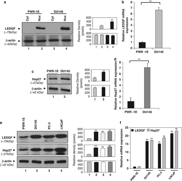
Figure 1.
LEDGF, a transregulator of Hsp27, and Hsp27 were aberrantly co-expressed in DU145 cancer cells. (a) Western analysis of LEDGF protein isolated from normal prostate epithelial cells, PWR-1E (upper panel, lanes 1 and 2) and human prostate carcinoma cell line DU145 (upper panel, lanes 3 and 4). Nuclear or cytosolic extracts from confluent cells were prepared. Equal amounts of protein were resolved through SDS-PAGE and immunoblotted using anti-LEDGF. Lower panel shows membrane probed with β-actin antibody as loading/internal control. Each band of blot was quantified using densitometer shown as histogram (right). Images are representatives from three independent observations. (b) mRNA level expression of LEDGF was analyzed by real-time PCR. Total RNA was isolated from PWR-1E (black bar) or DU145 (gray bar) cells and reverse transcribed. cDNA was subjected to real-time PCR analysis with specific primers. Data represent the mean±S.D. from three independent experiments (**P<0.001). (c and d) Expression levels of Hsp27 protein (c) and mRNA (d) in PWR-1E and DU145 cells. Data represent the mean±S.D. from three independent experiments (**P<0.001). (e and f) Expression levels of LEDGF and Hsp27 protein (e) and mRNA (f) in different prostate adenocarcinoma cell lines as indicated compared with normal prostate epithelium-derived cell line PWR-1E. Data represent the mean±S.D. from three independent experiments (**P<0.001). Each band of blot was quantified using densitometer shown at the right of western blot images

