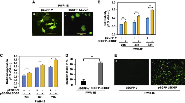
Figure 10.
Overexpression of LEDGF enhanced viability, proliferation and evoked invasive behavior of PWR-1E cells. (A) Representative images showing cells overexpressed with pEGFP-V or pEGFP-LEDGF in PWR-1E cells. (B) Histogram showing viability of PWR-1E cells overexpressed with pEGFP-LEDGF (yellow bars) compared with pEGFP-vector-transfected cells (blue bars) at time points shown (blue bars versus yellow bars). (C) Histogram showing proliferation of PWR-1E cells overexpressed with pEGFP-LEDGF (yellow bars) compared with pEGFP-vector-transfected cells (blue bars) at time points shown (blue bars versus yellow bars) using BrdU assay. Data represent the mean±S.D. from three independent experiments (**P<0.001). (D) Histogram showing the percentage of invasion index of PWR-1E cells overexpressing EGFP-LEDGF (gray bar). Data represent the mean±S.D. from three independent experiments (**P<0.001). (E) Photomicrographs of CyQuant GR-stained membrane with invaded cells (a, pEGFP-V; b, pEGFP-LEDGF). Images are of representatives from three independent experiments

