Figure 4.
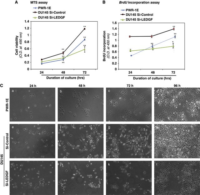
LEDGF depletion decreased the survival and proliferation of DU145 cells. (A) Graph showing the viability of Si-Control- and Si-LEDGF-transfected cells compared with PWR-1E normal prostate epithelial cells. Cells were harvested and processed for cell viability assay at different time points by MTS assay as depicted. (B) BrdU incorporation assay performed to evaluate the proliferation in LEDGF-depleted cells. Cells were labeled with BrdU for the last 2 h of culture and BrdU incorporation into DNA was measured by colorimetric assay followed by absorbance measurement at 450 nm as described by the company's protocol (Roche Diagnostics GmbH). Data represent the mean±S.D. from three independent experiments (**P<0.001; *P<0.01). (C) Representative photomicrographs of cultured cells attaining monolayer confluency (PWR-1E, a–d; Si-Control, e–h; Si-LEDGF, i–l). Images were taken at different time intervals as described in (C) with the aid of phase contrast microscope equipped with camera (Nikon Eclipse Ti-U phase contrast, Elgin, IL, USA)

