Figure 7.
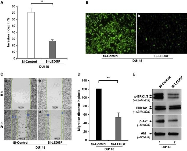
Knockdown of LEDGF attenuated the invasiveness and migration of DU145 cells and thereby deactivated ERK1/2/Akt signaling. (A) Cell invasion assay was carried out to evaluate the invasive behavior of cells after LEDGF depletion. Invasion index in percentage was calculated (open bar versus gray bar). Data represent the mean±S.D. from three independent experiments (**P<0.001). (B) The invaded cells on the membrane were stained with CyQunt GR dye and images were captured under FITC filter by Nikon Eclipse Ti-U fluorescent microscope equipped with camera. Images are representatives from three independent experiments (a, Si-Control; b, Si-LEDGF). (C) Images from wound-healing assay performed to assess the role of LEDGF in prostate carcinoma cell migration. Si-Control (a and c) and Si-LEDGF (b and d) cells were seeded in 60 mm petri plates and assay was performed. (D) Histogram showing the migration distance calculated from wound-healing assay (black bar versus gray bar). Data represent the mean±S.D. from three independent experiments (**P<0.001). (E) Western analysis performed to analyze the phosphorylation status of major MAPK kinases, ERK1/2 (upper panel) and Akt (third panel from the top). Total cell lysate from both Si-Control and Si-LEDGF cells were resolved on 10% SDS-PAGE and immunoblotted with anti-pERK1/2 or anti-ERK1/2 and anti-pAkt or anti-Akt antibodies. Level of phosphorylated and dephosphorylated forms of ERK1/2 and Akt are shown. Images are representatives from three independent experiments

