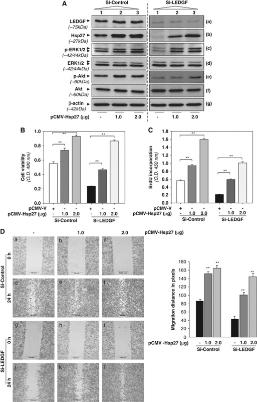
Figure 9.
LEDGF knockdown cells overexpressing Hsp27 reactivated ERK1/2 and Akt phosphorylation, regained its viability, proliferation and migratory behavior. (A) Representative images of western blot analysis performed for indicated proteins with cell lysates obtained from Si-Control (left panel) and Si-LEDGF (right panel) cells overexpressing Hsp27 of two different concentrations 1 μg (lane 2) and 2 μg (lane 3) of pCMV-Hsp27 or pCMV vector (lane 1)). Equal amount of plasmids were maintained with empty vector DNA in all transfections. The same membrane was used for striping and reprobed with different antibodies as indicated. Lower panel, β-actin band as internal control. (B and C) Histogram showing cell viability (B) and BrdU incorporation (C) of Si-Control and Si-LEDGF cells overexpressed with pCMV-Hsp27 compared with pCMV vector-transfected cells. Data represent the mean±S.D. from three independent experiments (**P<0.001). (D) Si-Control and Si-LEDGF cells overexpressing Hsp27 displayed higher migration activity. Wound-healing assay was performed in Si-sontrol and Si-LEDGF cells transfected either with pCMV-Hsp27 or pCMV vector. Images are representatives from three independent experiments. Upper panel, migration of Si-control cells transfected with either pCMV vector (a and d) or pCMV-Hsp27 (b, c, e and f). Lower panel, migration of Si-LEDGF cells transfected with either pCMV vector (g and j) or pCMV-Hsp27 (h, i, k and l). Migration distance was calculated using NIS-Elements BR 3.10 image analyzer software (Nikon) and is shown as a histogram (right panel, black bar versus gray bar). Data represent the mean±S.D. from three independent experiments (**P<0.001)

