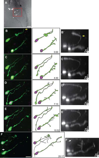
Figure 10. Time-lapse series of an immature neuron and developing radial glial cell.
24 hrs after transfection with pSox2-bd::turboRFPnls (magenta) and pSox2-bd::turboGFP (green), 5 complete confocal stacks were acquired over 25:17 hrs. A) Projection of the confocal stack of the right tectal lobe superimposed on a brightfield image of the tectal lobe to show tissue edges. Box indicates area of the cell shown in B-F. Scale bar = 50 μm. B-F) Cropped confocal projections of the cells and illustrations. B□-F□) 90° rotated projections of the same confocal stacks as in B-F. This perspective reveals the trajectory of the axon of the immature neuron (left) which grows up to the dorsal surface of the brain (yellow arrow) and then projects medially along of the pial surface. White arrow (F) indicates the tip of the axon. The glial cell (right) projects to the lateral edge of the brain and develops distinct endfeet over the course of the time-lapse (arrows). Scale bar = 20 μm.

