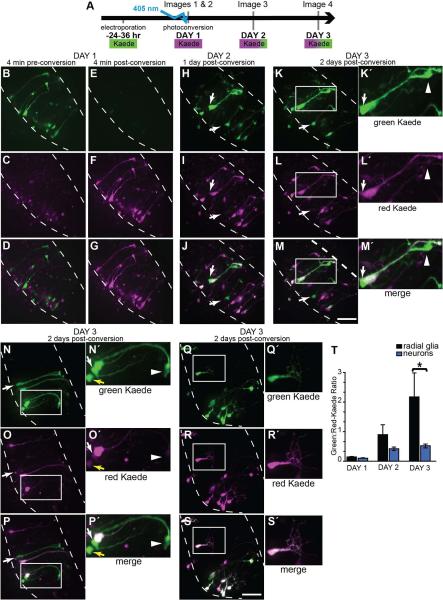
Figure 3. pSox2-bd::Kaede expression reveals newly born cells.
A) Diagram of the time-lapse imaging protocol used. 24 hrs after transfection with pSox2-bd::Kaede, complete confocal stacks were taken immediately before and after photoconversion. Two additional confocal stacks were acquired on each of the subsequent 2 days. Photoconversion of Kaede was achieved with ~20 sec. exposure of the tectal lobe with the 405 nm laser through the microscope objective. In proliferating cells, Kaede continues to be synthesized (appearing green), but cells generated after the photoconversion can be identified because they lack the photoconverted (magenta) Kaede. B-S) Flattened confocal stacks of the right tectal lobe of the unconverted green Kaede (top row), photoconverted red Kaede (magenta, middle row) and merged projections (bottom row). B-D) Kaede expression before and immediately after (E-G) photoconversion. The same tectal lobe on the 2nd (H-J) and 3rd day (K-M) where new, primarily green Kaede-expressing cells have appeared (arrows). K□-M□) Magnified views of boxes in K-M: A radial glial cell expressing high levels of unconverted Kaede. Arrows point to the cell body and the arrowhead at the pial endfoot of a radial glial cell that lacks photoconverted Kaede. It is closely apposed to a neighboring radial glial cell that expresses photoconverted Kaede. N-S) Kaede-positive cells in the right tectal lobe of two additional tadpoles imaged 2 days after photoconversion with an example of a newly-generated radial glial cell (boxed area and N□-P□) and neuron (boxed area and Q□-S□). Yellow arrow indicates the soma of an immature neuron. The white arrow points to the soma of a radial glial cell lacking photoconverted Kaede. Its pial endfoot is shown with the arrowhead. T) Quantification of the red- and green-fluorescent signals from pSox2-bd::Kaede-expressing cells beginning immediately after photoconversion for 3 consecutive days. The increasing green:red ratio of Kaede expression reveals that radial glial cells express significantly greater levels of Sox2 than neurons. MW, p = 0.01. Intensity values for red and green Kaede fluorescence are reported in Table 2.

