
Figure 5. Prolonged time course of cell division in vivo.
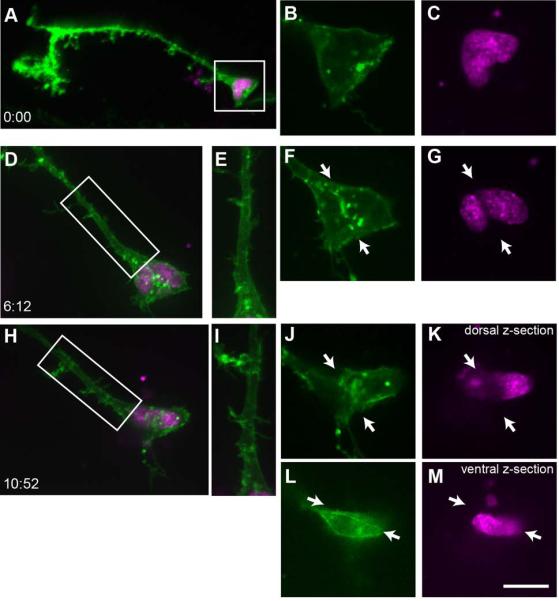
A) A projection of a tectal radial glial cell expressing membrane-targeted GFP (pSox2-bd::mGFP, green signal) and pSox2-bd::RFPnls (magenta) reveals the complex morphology of the pial endfoot, many fine filopodia extending from the radial process and endfoot, and a lobed nucleus. B-C) Enlarged single z-planes through the cell body captured at the same time point as in A. At the first time point, the nucleus is dimpled but not divided (C) and there is no evidence of mGFP-labeled plasma membrane in the center of the cell body between the nuclear lobes (B). D-G) ~6 hours later, the nuclei have separated and mGFP-labeled membrane extends between the nuclei (arrows, F, G). Only one radial process is visible (boxed region of D is enlarged in E). By the third time point, almost 5 hours later (H-M), the nuclei have rotated so they are above and below one another, and remain separated by GFP-labeled membrane (arrows, J-M). Because of the dorso-ventral orientation of the nuclei, two Z-planes separated by ~7 μm are shown: a more dorsal plane (J, K) and ventral plane (L, M). There is no evidence of a second radial process extending from the newly generated cell (boxed region of H is enlarged in I).