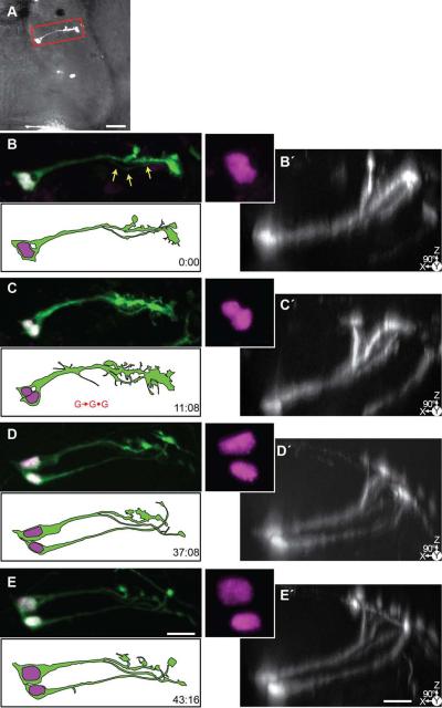
Figure 9. Time-lapse series of a symmetrically dividing radial glial cell.
24 hrs after transfection with pSox2-bd::turboRFPnls (magenta) and pSox2-bd::turboGFP (green), 4 complete confocal stacks were acquired over 43:16 hrs. A) A brightfield image of the tectal lobe overlaid with the projection of the confocal stack of the tectum cropped in Z around the cell shown in B-E. Scale bar = 50 μm. B-E) Projections of the cropped confocal stacks of the radial glial cell, and same stacks (B□-E□) rotated 90° to reveal the dorsal reaching pial endfeet of the glia in the Z-dimension. Note the presence of a faint process of the second cell beginning at t = 0:00 (arrows). The cell divides and nuclei separate (C, marked with G→G•G). At the final time point, two glia, each with endfeet, are visible. Scale bar = 20 μm.

