
Figure 19.
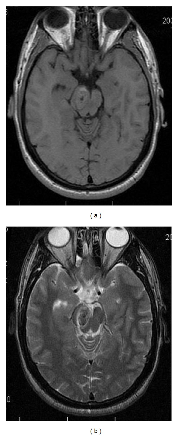
Benedikt Syndome. Ovoid lesion within the midbrain demonstrates isointensity on axial T1-weighted image (a), and slight hypointensity on axial T2-weighted (b) image. These findings are consistent with a cavernous hemangioma within the right midbrain, presenting with symptoms of Benedikt Syndrome.