
Figure 20.
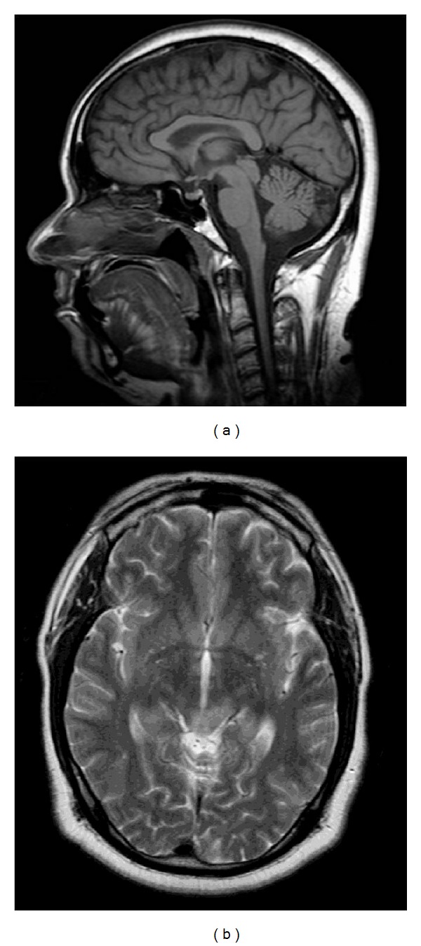
Nothnagel Syndrome. MRI images of the midbrain with enlargement of the midbrain tectum, including the quadrageminal plate, noted on sagittal T1-weighted (a) image. There is associated abnormal T2 hyperintensity seen on accompanying axial T2-weighted (b) image. These findings likely represent tectal glioma with involvement of the oculomotor nuclear complex and decussating fibers of the superior cerebellar peduncle.