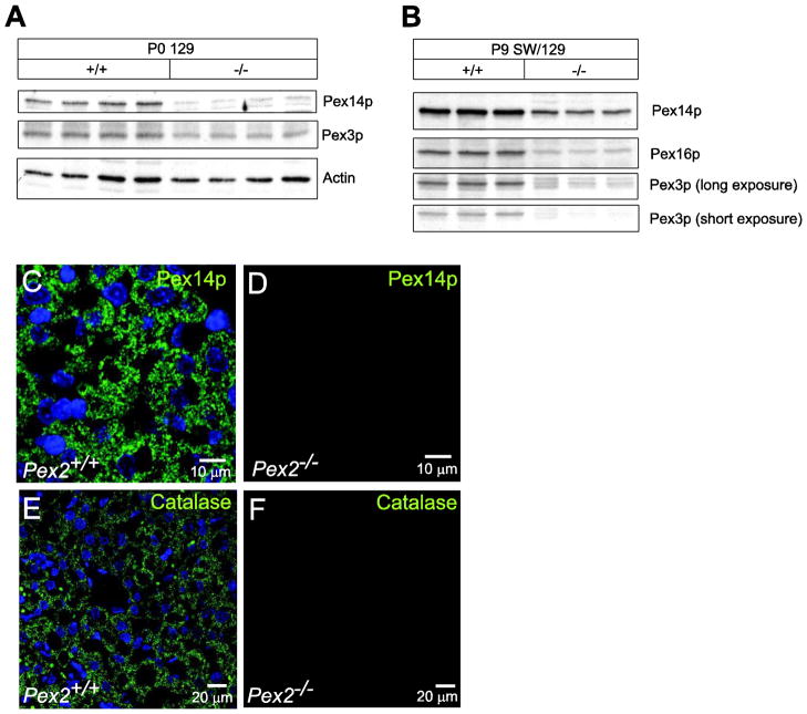
Figure 6. Immunoblot and immunofluorescence analysis of peroxin proteins and catalase in livers from control and Pex2−/− mice.
(A, B) Protein lysates from livers of newborn 129 (A) and 9-day-old SW/129 (B) control and Pex2−/− mice were probed by immunoblot as labeled. (C–F) Liver sections from P9 wild-type (C, E) and Pex2−/− (D, F) mice were stained with an antibody to Pex14p (C, D) and catalase (E, F) and imaged by confocal microscopy. Peroxisomes (C) and peroxisome membrane ghosts (D) were detected using an antibody to Pex14p. Note that the number of peroxisome membrane ghosts in Pex2−/− mice is significantly lower than the number of peroxisomes in wild-type mice. PTS1 protein import was assessed as the distribution of punctate (organelle-bound) (E) versus cytoplasmic (F) catalase. Scale bar: 10 μm for panels C, D; 20 μm for panels E, F.

