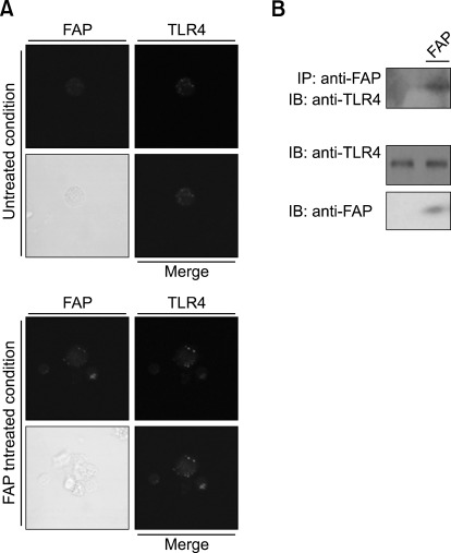
Figure 1.
FAP interacts with TLR4. (A) Co-localization of FAP and TLR4 on DCs. DCs were treated with FAP (500 ng/ml) for 30 min, fixed, and stained with rabbit anti-FAP and mouse PE-conjugated anti-TLR4 antibodies overnight at 4℃, and then stained with Alexa568-conjugated anti-rabbit antibody for 1 h at room temperature. Cell morphology and fluorescence intensity were analyzed using the Zeiss LSM510 Meta confocal laser scanning microscope. (B) Co-immunoprecipitation of FAP and TLR4. DCs were treated with FAP (500 ng/ml) for 30 min. Cells were harvested, cell lysates were immunoprecipitated with IgG and anti-FAP, and proteins were visualized by immunoblotting with an anti-TLR4 antibody.

