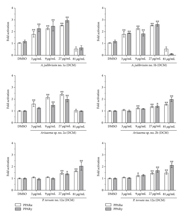
Figure 1.

Dose-response experiments with DCM extracts of CHMs of A. julibrissin no. 1a and b, Arisaema sp. no. 2a and b, and P. ternata no. 12a and b. HEK293 cells were transiently transfected with the expression plasmid for PPARα or PPARγ, the reporter plasmid pPPRE-tk3x-luc, and the internal control plasmid (EGFP). The transiently transfected cells were incubated for 18 h with 3, 9, 27, and 81 μg/mL of each indicated extract. Furthermore, the cells were similarly incubated with 0.1% DMSO (negative control), 50 nM GW7647 (PPARα agonist, activated PPARα 2.5–3.5 fold but not PPARγ), or 5 μM troglitazone (PPARγ agonist, activated 3.5–6.5 fold PPARγ but not PPARα) as positive controls (not shown). Luciferase activity and fluorescence intensity were measured. Results are presented as mean ± SD, (n = 4). Significantly different from the negative control (ANOVA), *P < 0.05; **P < 0.01.
