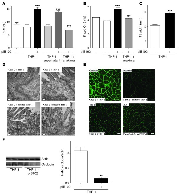
Figure 7. IL-1β secreted by Y. pseudotuberculosis–infected THP-1 cells alters paracellular permeability and E. coli translocation across Caco-2 monolayers.
(A) Caco-2 and (B) Caco-2 clone-1 cells were cultivated into TCs. Then, THP-1 cells infected or not with pIB102 were added to the TC basolateral compartment, and (A) paracellular permeability and (B) E. coli translocation were monitored. To investigate the involvement of IL-1β receptor, Caco-2 and Caco-2 clone-1 cells were treated for 24 hours with anakinra (50 μg/ml). n ≥ 10 per group; 3 independent experiments. ***P < 0.001 versus basal (–); †††P < 0.001 versus pIB102-infected THP-1. (C and D) TJ width of Caco-2 cells incubated with infected THP-1 cells was measured by EM. Scale bars: 100 nm. n = 150 measures of TJs per group from 3 independent wells. ***P < 0.001 versus uninfected THP-1. (E) Apical distribution of occludin and ZO-1 of Caco-2 cells incubated with infected THP-1 cells was analyzed by confocal microscopy. Scale bar: 20 μm. (F) Western blot analysis of occludin in Caco-2 cells incubated with infected THP-1 cells. n = 3 per group. **P < 0.01 versus uninfected THP-1.

