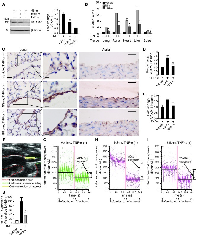
Figure 2. miR-181b represses TNF-α–induced proinflammatory gene expression in vivo.
(A) Mice were i.v. injected with vehicle, miRNA negative control, or miR-181b mimics (50 μg/mouse). Twenty-four hours later, mice were treated with or without TNF-α for 4 hours, and lungs were harvested for Western blot analysis of VCAM-1 protein levels. Densitometry was performed and fold change of protein expression was quantified. (B) Experiments were carried out as described in A, and real-time qPCR analysis of VCAM-1 mRNA level in indicated tissues was performed. (C) VCAM-1 staining of lung and aorta sections. Mice were treated as in A. Scale bars: 25 μm (insets, 10 μm). (D and E) Quantification of VCAM-1 staining in lung and aortic endothelium, respectively. (A–E) Vehicle group (n = 3 mice), miRNA negative control group (n = 5 mice), miR-181b mimics group (n = 5 mice). Data represent mean ± SEM. (F) Ultrasound image shows region of interest (innominate artery) for in vivo VCAM-1 imaging using microbubble contrast. (G–I) Mice were injected with vehicle, miRNA negative control (n = 7), or miR-181b mimics (n = 6). Representative images show the differential targeted enhancement values for VCAM-1 expression detected by ultrasound before and after microbubble burst. (J) Quantification of differential targeted enhancement values for VCAM-1 expression in mice injected with miRNA negative control or miR-181b mimics. Data represent mean ± SEM. *P < 0.05.

