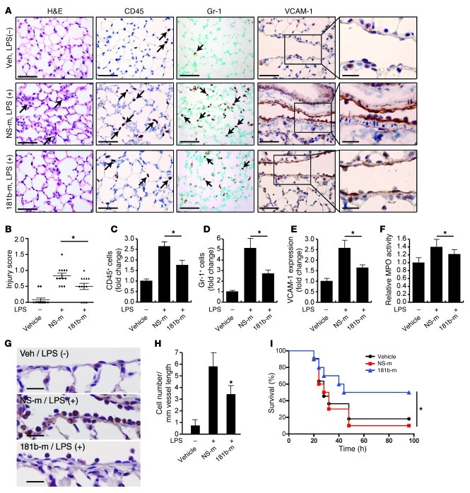
Figure 6. miR-181b reduces EC activation and leukocyte accumulation in LPS-induced lung inflammation/injury.
(A) Mice were i.v. injected with vehicle, miRNA negative control, or miR-181b mimics and treated with or without LPS (40 mg/kg, i.p., serotype 026:B6) for 4 hours; lungs were harvested and stained for H&E, Gr-1, CD45, or VCAM-1 staining. Scale bars: 50 μm (insets, 20 μm). (B) Evaluation of lung injury 4 hours after LPS was determined by lung injury scoring. Each data point represents score from 1 section. n = 4 mice per group, and 3 sections per mouse were scored. *P < 0.05. (C) Quantification of CD45-positive cells. *P < 0.05. (D) Quantification of Gr-1 positive cells. *P < 0.05. (E) Quantification of VCAM-1 expression. *P < 0.05. n = 4 mice per group; values represent mean ± SD (C–E). (F) Mice were treated as in A. Lungs were harvested and assessed for MPO activity, and the value of the vehicle group was set to 1. Values represent mean ± SD, n = 6 mice per group. (G and H) Mice were treated as in A, and lungs were harvested for Gr-1 staining. Scale bars: 20 μm. Quantification shows the number of Gr-1–positive cells per mm vessel length. Values represent mean ± SD, n = 4. *P < 0.05. (I) Kaplan-Meier survival curves of: LPS-treated C57BL/6 mice (50 mg per kg, i.p., n = 10 to 11 per group) that were injected i.v. with vehicle (black circles), miRNA negative control (red squares), or miR-181b mimics (blue triangles) 48 hours before, 24 hours before, and 1.5 hours after LPS administration. *P < 0.05, 1-tailed log-rank test.

