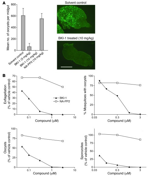
Figure 3. BKI-1 blocks formation of oocysts and thus malaria infection of mosquitoes.
(A) Mice were infected with GFP-expressing P. berghei and treated with compounds at 3 mg/kg or 10 mg/kg i.p., and 30 minutes later, mosquitoes were fed on mice. Shown are the geometric mean oocyst number on 8–20 dissected mosquito midguts explanted 5 days after feeding. Fluorescence micrographs illustrate typical infection levels in mosquitoes fed on control and BKI-1–treated mice. Scale bar: 500 μm. Data are representative of 3 experiments. Error bars represent SEM. (B) Compounds were mixed with human blood and aliquots explanted for exflagellation observation, and the blood was fed to A. stephensi mosquitoes. A complete suppression of exflagellation was observed with 1 and 3 μM BKI-1. Blocking exflagellation with BKI-1 correlated well with the prevention of oocyst and infective sporozoite formation. Sexual stage development in mosquitoes fed with 0.1 μM was not completely but was significantly reduced, as shown by more than 86% in the number of oocyst and infective sporozoites. In a repeat experiment, exflagellation events in the presence of BKI-1 were suppressed completely at 1 or 3 μM and more than 90% at 0.3 μM.

