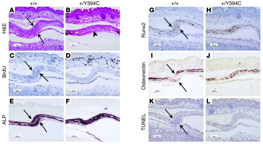
Figure 3. Fgfr2+/Y394C mice show abnormal histology, proliferation, and differentiation in the skull.
(A and B) H&E staining shows the abnormal development of the mutant coronal suture at E17.5, with presynostosis and osteoid deposition between the osteogenic fronts (arrows, osteogenic fronts; arrowhead, presynostosis/synostosis). (C and D) Immunohistochemical staining of BrdU shows decreased numbers and abnormal distribution of positive cells in mutants at E17.5. (E and F) ALP staining shows broad and expanded ALP expression into the coronal suture of the mutants at E17.5. (G and H) Immunohistochemical staining of Runx2 shows increased expression and abnormal differentiation at the osteogenic fronts at E17.5. (I and J) In situ hybridization of osteonectin shows accelerated bone formation in mutants at E17.5. (K and L) TUNEL staining shows no obvious change of apoptosis in the mutant. A, C, E, G, I, and K are from littermate controls. B, D, F, H, J, and L show the corresponding organs and tissues in mutant mice. Scale bars: 50 μm.

