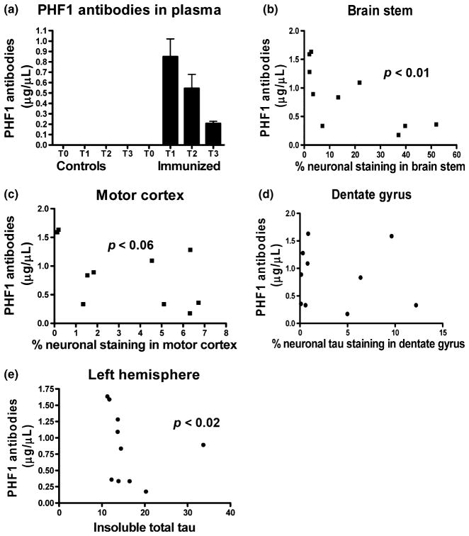
Fig. 4.
(a) PHF1 antibodies were cleared relatively quickly from plasma. No detectable antibodies were observed in controls, whereas the levels in immunized animals decreased over time. The rate of clearance appeared to be faster than for endogenous IgG (typical half-life of IgG is 21–28 days), as is often observed for therapeutic monoclonals (Keizer et al. 2010). Each bar represents the average values for the immunized mice + SEM. T0: prior to first immunization, T1: 24 h after the 12th injection, T2: 7 days after the 13th and last injection, T3: 14 days after last injection. The ELISA plates were coated with Tau379–408[P-Ser396, 404]. n = 10 per group per each time point. (b–e) Plasma levels of PHF1 antibodies correlated inversely with tau pathology. Significant correlation was observed in the brainstem (b; p < 0.01), and a strong trend for correlation in the motor cortex (c; p = 0.06). A similar pattern, albeit with an outlier, was observed in the dentate gyrus (d). Similar pattern was detected when antibody levels were compared with levels of insoluble total tau on western blots (e; p < 0.02). n = 10 per group.

