Figure 5.

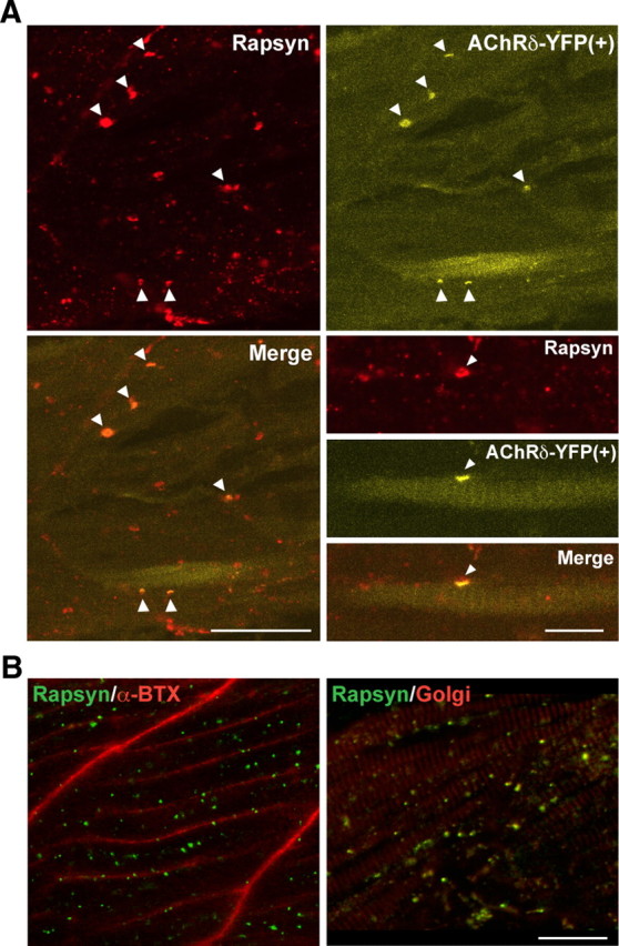
Rescue of rapsyn transport by muscle-type AChRs. A, Expression of AChRδ-YFP in sofa potato mutants restored the membrane distribution of rapsyn. In muscle cells expressing YFP, the distribution of rapsyn visualized by antibody (top left) and that of AChR visualized by YFP (top right) colocalized (arrowheads). The merged image is shown in the bottom left. Note that only a subpopulation of muscle cells expressed AChRδ-YFP and formed coclusters of AChR/rapsyn. Scale bar, 50 μm. Higher-magnification pictures of a single muscle cell expressing YFP are shown in the bottom right. Rapsyn and AChR (arrowheads) are colocalized on the plasma membrane. Scale bar, 20 μm. B, A sofa potato larva expressing α7-YFP in all muscle cells. While α7-YFP is detected on the plasma membrane (α-Btx shown in red; left), rapsyn (green) does not colocalize with α7-YFP but colocalizes with the Golgi marker (GM-130 shown in red; right). Note that two panels are from different larvae. Scale bar, 20 μm.
