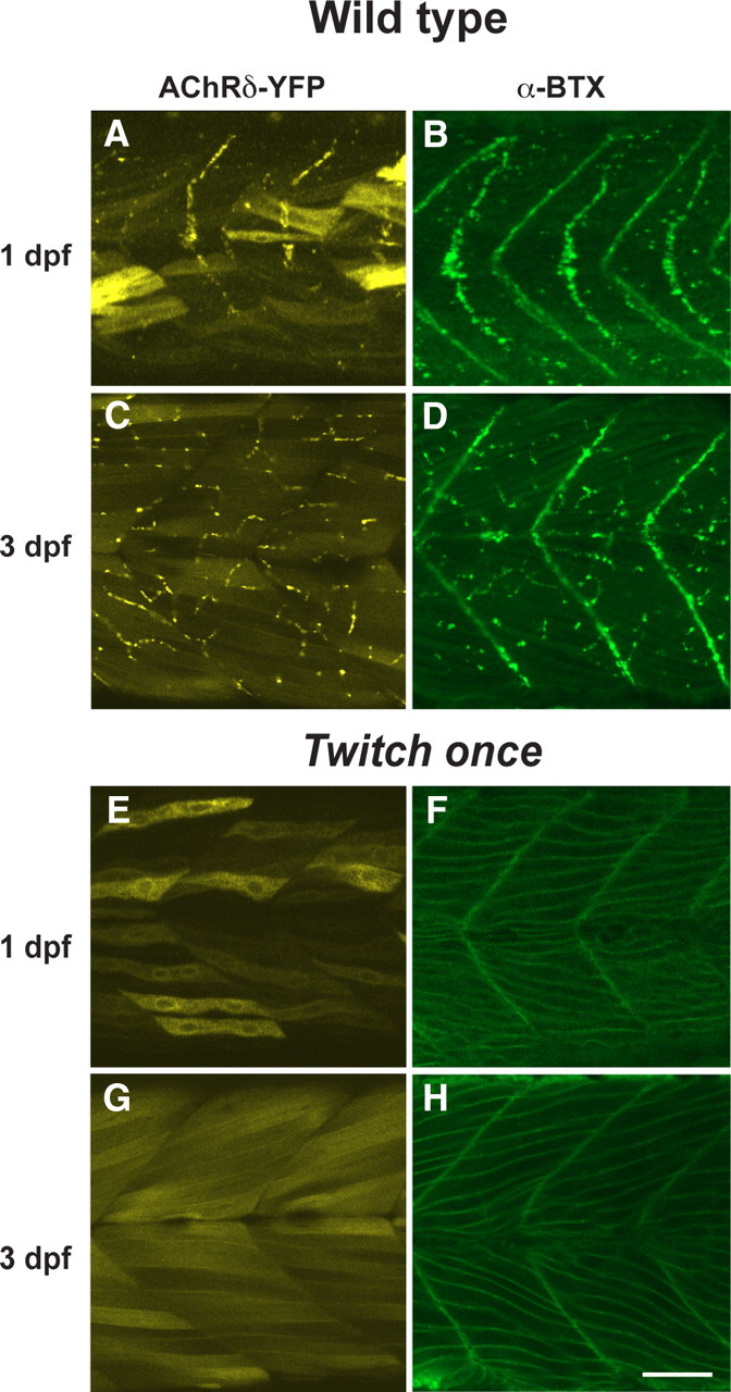
Figure 7.

The distribution of AChR was observed in AChRδ-YFP(+) larva with YFP (A, C, E, G) and in native larvae stained with α-Btx (B, D, F, H) in the wild-type (A–D) or the twitch once background (E–H). While the YFP molecule filling the cytoplasm obscures the AChRδ-YFP distribution in twitch once (E, G), the membranous distribution of AChRs is obvious with α-Btx (F, H), which stains only assembled pentamers. Scale bar, 50 μm.
