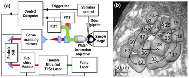
Fig. 5.
a Schematic setup of the two-photon microscope: A tunable ultra-short pulsed laser (Mai Tai Deep See HP, Spectra-Physics) is dispersion-compensated in pre-chirp module. A Pockels cell controls the light intensity, and galvo-mirrors allow for fast and variable scanning. The beam is strongly focussed onto the sample by a water immersion objective (40×, NA 0.8, Olympus). Fluorescence is collected by the same objective, separated from the backscattered excitation light by a dichroic beam-splitter, split into green and red detection channels by dichroics and band-pass filters (Chroma Technology), and detected by Photomultiplier tubes (PMT, Hamamatsu Photonics). A computer controls all microscope parameters and synchronises imaging with a odour stimulus generator. b Axial projection view of a right AL in A. mellifera forager, reconstructed volume images of a subset of T1 glomeruli (labelled according to Galizia et al. 1999b); the black spiral represents the custom-defined scan trace for fast functional imaging of the glomerular activity (reprinted from Haase et al. 2011 with permission from OSA)

