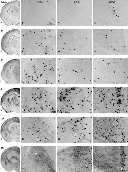
Figure 2.
Histological sections from P7 mouse brain treated with saline (A–D) or PG (E–X) at various time points after exposure. Black arrows depict AC-3 positive-cells. Hemi-sections are taken from the same rostro-caudal level (10× magnification). High-powered photomicrographs (20× magnification) are taken from the CPu (*), LD Th (†), and retrosplenial cortex (RSC; §), as indicated by a symbol in panel A. Onset of damage appeared at 2h in the CPu (F). Peak levels of damage occurred at 8h in most regions (M–P). Overall AC-3 staining was decreased at 12h (Q–T) and 24h (U–X), where staining was specific to intact cell bodies (white arrow heads). In addition to localized staining, a non-specific diffuse staining was present at 24h.

