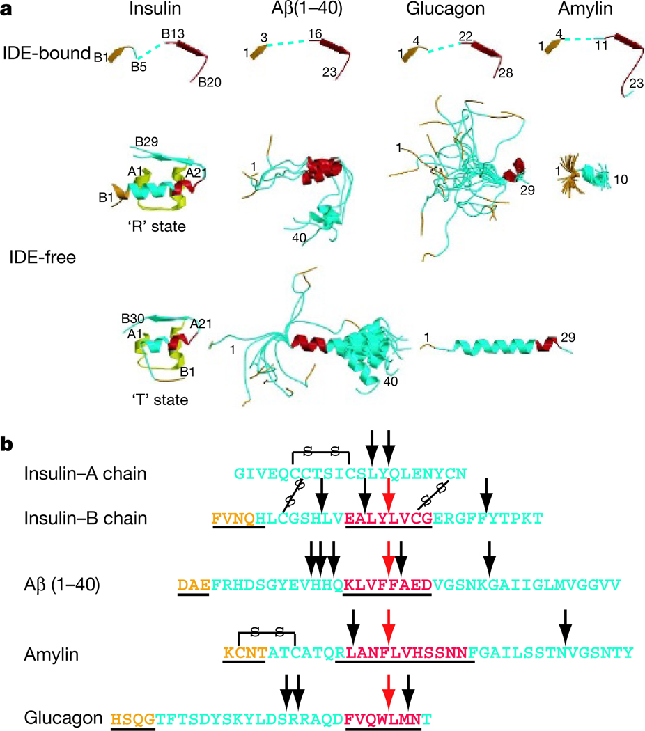
Figure 4. Conformational changes and catalysis of IDE substrates.
a, Secondary structure of IDE substrates in the IDE-bound (top) or free (bottom) form. The N terminus and IDE catalytic cleft binding segment are coloured orange and red, respectively. The PDB accession codes for insulin are 1G7A and 1ZEH, those for Aβ are 1AML and 1BA4, those for glucagon are 1GCN and 1KX6, and that for amylin is 1KUW. b, Sequence comparison of four IDE substrates. Arrows indicate the main cleavage sites of the substrate by IDE1,26 (Supplementary Fig. 13). Amino acids that are underlined are observed in the crystal structures of substrate-bound IDE.

