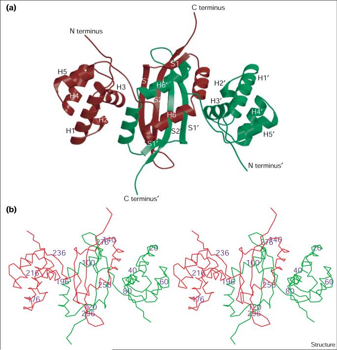
Figure 2.
The cyanase dimer. (a) Ribbon diagram of the cyanase dimer. The cyanase monomers are colored red and green. Secondary structure elements are labeled H for an α helix and S for a β strand: helix 1 (H1) consists of residues 8–24; helix 2 (H2), 29–33; helix 3 (H3), 40–47; helix 4 (H4), 55–64; helix 5 (H5), 69–76; helix 6 (H6), 92–115; strand 1 (S1), 119–134; strand 2 (S2), 140–152. A prime symbol distinguishes the two monomers. (b) Stereoview Cα trace of the cyanase dimer. The colors are as described in (a) with every 20th residue labeled. (The figure was generated with Molscript [62] and rendered with Raster 3D [63].)

