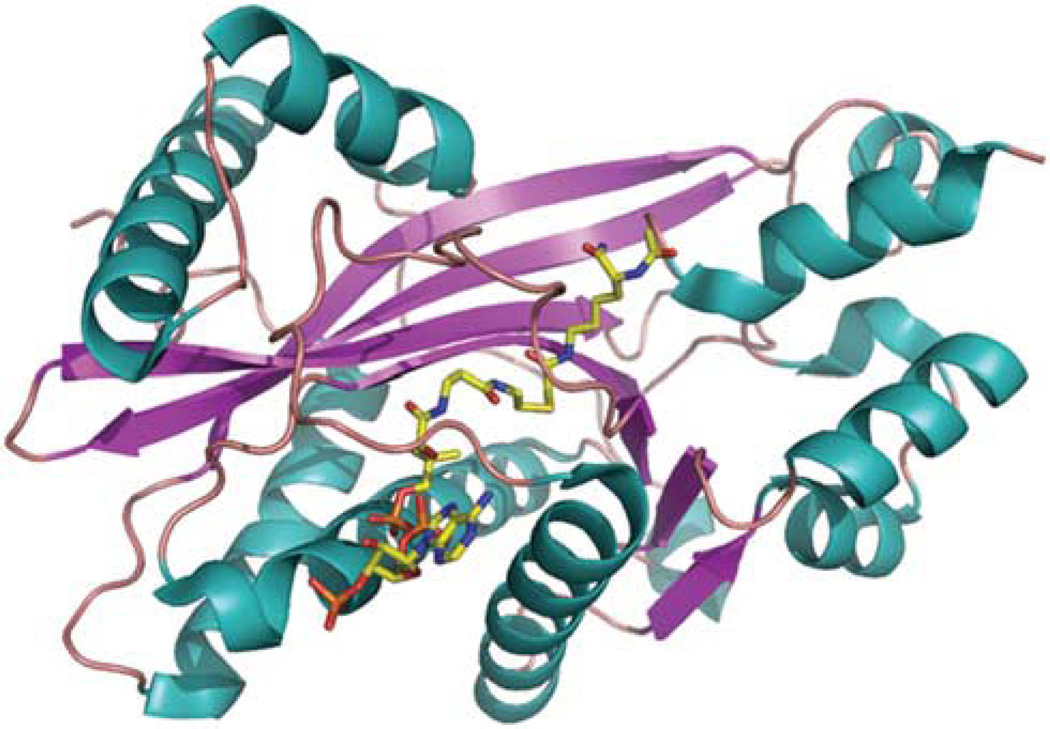
Figure 3.
Structure of p300 HAT domain. Crystal structure of p300 HAT domain (PDB code: 3biy) was solved in complex with a bisubstrate inhibitor, Lys-CoA.61 Two HAT subdomains, amino acids 1287–1522 and amino acids 1569–1666, were co-purified and used for crystallization. The structure is showed as cartoon and colored by secondary structure (alpha helix: teal, beta strand: magentas; loop: salmon). The inhibitor in the structure is presented as sticks and colored by atomic type (carbon: yellow; nitrogen: blue; oxygen: red; phosphorus: orange).

