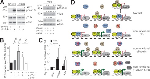
FIGURE 6.
Cells with reduced expression of γ-tubulin and RB1 undergo apoptosis. A, Y79, U1690, or U2OS cells were transfected with control-shRNA (shcont.), γ-TUBULIN-shRNA (shγTub), or E2F1-shRNA (shE2F1) vectors; after 48 h, total lysate from 1 × 10⁶ were prepared and examined by Western blotting (WB) using antibodies against pro-caspase 3 (pcasp.3), E2F1, γ-tubulin, and α-tubulin (Tub) (n = 3). Numbers above Western blots indicate variations on protein expression relative to control. To adjust for differences in protein loading, the protein concentration of RB1, E2F1, and γ-tubulin was determined by their ratio with α-tubulin for each treatment. The protein ratio in control treatment was set to 1. B and C, aliquots of different cell lines (as indicated), treated as in A for 72 h, were stained with annexin V-FITC and 7AAD and analyzed by flow cytometry (B) or stained with trypan blue and analyzed by morphology (C). The number of annexin V- and 7AAD-positive cells transfected with control construct was set as 1, and relative changes were calculated (mean ± S.D. (error bars), n = 3–6). D, γ-tubulin (γTub) and RB1 (RB) protein levels regulate each other's expression by direct binding to respective promoters. From left to right along each line RB1, TUBG1, and caspase 3 (CASP3) genes are represented with their respective promoter regions. Red lines and green arrows represent repressed and activated genes, respectively. Red cross denotes the absence of protein expression of the represented protein. In a cell, the presence of γ-tubulin and RB1 represses transcription from the RB1 and TUBG1 genes, respectively. The absence of RB1 increases the transcriptional activity of the TUBG1 gene, and accordingly, the absence of γ-tubulin increases the transcriptional activity of the RB1 gene. Finally, concomitant reduced levels of γ-tubulin and RB1 will lead to increase expression of CASP3. In promoter regions of repressed genes, RB1 is in complex with the transcription factor heterodimer E2F1-Dp. Alternatively, the inhibiting protein complex could be E2F–γ-tubulin. Black arrows point toward the two possible protein complexes that may repress expression of the CASP3.

