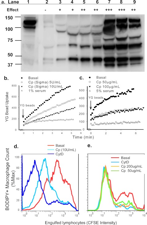
FIGURE 3.
CP inhibits phagocytosis of beads and apoptotic cells. a, immunoreactive CP protein in column fractions was detected by Western blotting. Lane 1, starting material; lanes 2∼9, fractions (30 μl each) eluted from Superdex 200 gel filtration. The relative inhibitory effect of each fraction on YG bead uptake by monocytes is indicated (−, no inhibition; + to +++, increasing strength of inhibition). b and c, human PBMC labeled with APC-conjugated anti-CD14 mAb were resuspended in sodium medium with 0.1 mm Ca2+. CP or serum was added 1 min before the addition of YG beads. The fluorescence intensity of CD14+ monocytes containing engulfed beads were analyzed by time-resolved flow cytometry. d and e, flow cytometry histograms show phagocytosis of CFSE-labeled apoptotic lymphocytes by autologous monocyte-derived macrophages labeled with CMTMR or BODIPY. Mixed cells were incubated with CP or CytD (20 μm) for 3 h before cells were collected and fixed for flow cytometry assay. Inhibitors used were commercially obtained CP (∼50 unit/mg) (b and d), CP purified from human serum (c and e), or human serum (b and c).

