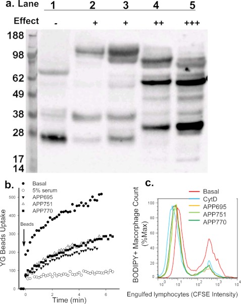
FIGURE 5.
APP inhibits phagocytosis of beads and apoptotic cells. a, shown is APP protein expression detected by Western blotting with anti-APP mAb (clone 22C11). Lanes 1∼5, fractions eluted from Superdex 200 gel filtration. The relative inhibitory effect of each fraction on YG bead uptake by monocytes is indicated. b, human PBMC labeled with APC conjugated anti-CD14 mAb were resuspended in sodium medium with 0.1 mm Ca2+. Recombinant APP695α, APP751α, or APP770α (100 μg/ml each) was added 1 min before the addition of YG beads. The uptake of YG beads by CD14+ monocytes were analyzed by time-resolved flow cytometry. c, flow cytometry histograms show phagocytosis of CFSE-labeled apoptotic lymphocytes by autologous monocyte-derived macrophages labeled with CMTMR or BODIPY. Mixed cells were incubated with isoforms of APP (200 μg/ml) or CytD (20 μm) for 3 h before cells were collected and fixed for flow cytometry assay.

