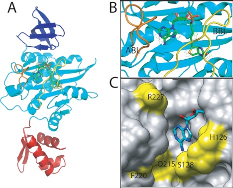
FIGURE 2.
S. aureus BPL in complex with biotinol-5′-AMP. A, SaBPL consists of three structured domains, an N-terminal DNA binding domain (red), a central domain (cyan), and C-terminal domain (dark blue). B, a close-up of the inhibitor binding site shows the relative positions of the ABL (orange) and BBL (yellow). The final 2Fo − 2Fc map is contoured at the 1σ level is shown on inhibitor 2 in ball and stick representation. C, the ATP pocket of SaBPL in complex with 2 is shown in space-filled mode with the adenine portion shown in ball and stick representation. Amino acid residues that line the pocket and that are not conserved between SaBPL and human BPL are highlighted in yellow.

