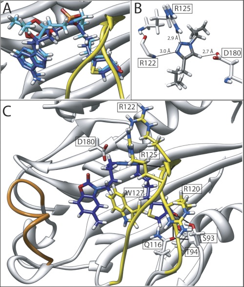
FIGURE 5.
Mode of inhibitor binding. A, the backbone atoms of SaBPL in complex with inhibitor 2 (dark blue) and inhibitor 7 (cyan) were superimposed to reveal the remarkable overlap in the conformations imparted by the triazole bioisostere. B, hydrogen bonding interactions between SaBPL and the trizole ring are shown. C, the crystal structure of biotin triazole 14 (dark blue) bound in the active site of SaBPL. Hydrogen bonding contacts with the amino acids of SaBPL are shown. The BBL is highlighted in yellow, and the ABL is in orange.

