FIGURE 7.
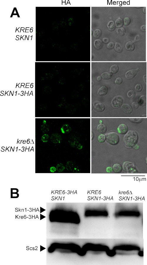
Immunofluorescence images and amounts of Skn1-3HA protein in the presence or absence of its homologue Kre6. A, the epitope-tagged Skn1-3HA was expressed from the original chromosomal SKN1 locus in either KRE6 or kre6Δ background and detected by indirect immunofluorescence staining with mouse anti-HA monoclonal antibody (HA, left panels). The Nomarski images are merged to show the cells (Merged, right panels). The images of wild-type KRE6 SKN1 cells were included to show the background signals without HA epitope. The bar indicates 10 μm. B, the same amount of lysates from KRE6-3HA SKN1, KRE6 SKN1-3HA, or kre6Δ SKN1-3HA cells were subjected to SDS-PAGE and immunoblotting using anti-HA antibody. The immunoblotting bands of Scs2 are the loading control.

