FIGURE 3.
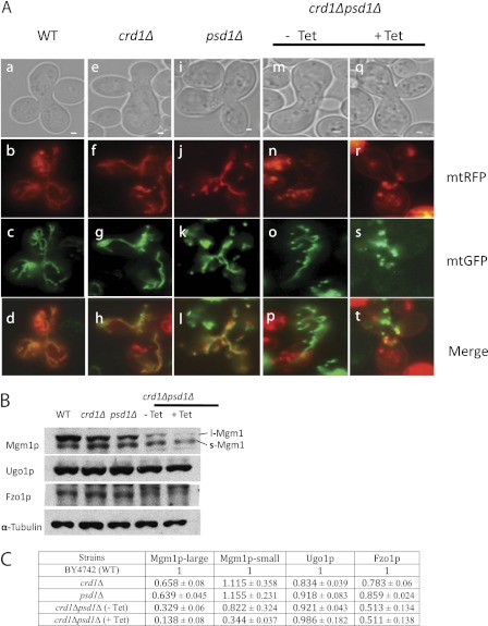
crd1Δpsd1Δ cells exhibit defective mitochondrial fusion. A, cells of opposite mating types were transformed with either mtGFP or mtRFP. Mitochondrial fusion was examined by observing merged images of mtGFP and mtRFP in WT (a–d panels), crd1Δ (e–h panels), psd1Δ (i–l panels), and crd1Δpsd1Δ cells grown without (m–p panels) or with (q–t panels) tetracycline (Tet). Bars, 1 μm. B, total cellular proteins were analyzed by SDS-PAGE followed by Western blot. Steady state levels of Mgm1p, Fzo1p, and Ugo1p were measured. α-Tubulin was used as a loading control. C, quantitation of fusion proteins. Values are mean ± S.E. (n = 3).

