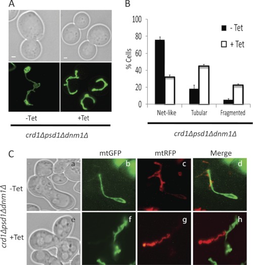
FIGURE 5.
crd1Δpsd1Δdnm1Δ cells are defective in mitochondrial fusion. A, mitochondria were visualized in the crd1Δpsd1Δdnm1Δ mutant using mtGFP. Cells were grown at 30° C to log phase in synthetic deficient glucose medium with 200 μg/ml tetracycline (Tet) where indicated and examined by fluorescence microscopy. Bars, 1 μm. B, cells containing tubular, fragmented, and net-like mitochondria were quantified. Values are mean ± S.E. (n = 3). At least 100 cells were visualized in each experiment. C, crd1Δpsd1Δdnm1Δ cells of opposite mating types were transformed with either mtGFP or mtRFP. Mitochondrial fusion was examined by observing merged images of mtGFP and mtRFP in zygotes of crd1Δpsd1Δdnm1Δ grown without (a–d panels) or with (e–h panels) tetracycline. Bars, 1 μm.

