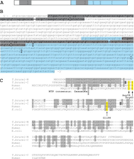
FIGURE 2.
DNA and amino acid sequences of T. brucei TK. A and B, schematic figure (A) and DNA sequence (B) of the T. brucei TK ORF with its two homologous domains shown in white and blue. Nearly identical 89-bp fragments present in the two domains are highlighted in gray (the only base that differs between the two fragments is not highlighted). The paired nucleotides (shown as one on top of the other) in A and B represent differences in the sequence between the two T. brucei TC221 alleles, TKallele 1 (top bases) and TKallele 2 (bottom bases). C, amino acid sequence alignment of T. brucei TKallele 1 N- and C-terminal parts (domains 1 and 2) with human and E. coli TK1 proteins. The human numbering scale is used. Amino acids conserved in the two T. brucei TK domains are shown in light gray, and the conserved amino acids that are substituted in domain 1 are highlighted in yellow. The two dark gray residues, A and G, are replaced by T and D in TKallele 2.

