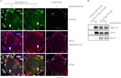
FIGURE 2.
Localization of Siglec-15 in the osteoclast plasma membrane. A, BMMs were cultured with RANKL and M-CSF for 96 h and subjected to immunostaining with the anti-Siglec-15 antibody with or without cell permeabilization. Normal rabbit IgG was used as a control antibody. Green, antibody-related signals; red, wheat germ agglutinin (WGA)-rhodamine; blue, Hoechst 33342. Arrowheads indicate the plasma membranes of multinucleated osteoclasts. B, cell surface biotinylation analysis. BMMs were cultured as described in A and treated with or without the Sulfo-NHS-SS-Biotin. Cell lysates were prepared, and biotinylated proteins were purified with avidin beads, and elution fractions from 62.5 μg of total cell lysates from mock-treated and biotin-labeled cells were loaded in the left two lanes. Total cell lysates (6.25 μg) before the avidin beads were loaded in the right two lanes to confirm that equal amounts of cell extracts were used in the avidin-bead purification step. c-Fms and Erk1/2 were used as control plasma membrane and cytosolic proteins, respectively. The asterisk indicates nonspecific signals as described for Fig. 1, B and C.

